SBPMD Histology Laboratory Manual
Prostate Gland
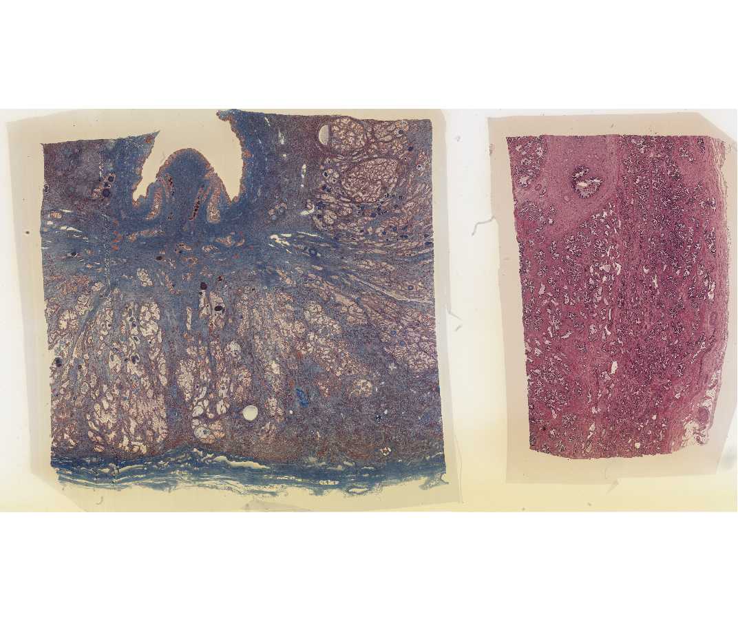
#59 Prostate, Human Age 55; 2 sections: Masson, and H&E
Open with WebViewer
Examine with the naked eye, studying particularly the Masson-stained section. At one surface, a prominent fold represents the cut surface of the prostatic urethra. Also evident are the elongate tubules forming the parenchyma of the gland, and also the dense fibrous connective tissue capsule. Locate the region of the prostatic urethra with the microscope, and study its lining epithelium. Compare its transitional epithelium with the epithelium lining the ducts and glands of the prostate, which can be cuboidal, columnar or pseudostratified. The tubulo-alveolar glands of the prostate are embedded in an abundant stroma of fibro-elastic connective tissue, which is interlaced, with strands of smooth muscle. Numerous concretions (corpora amylacea) occur in the lumen of the glands and ducts, and these tend to increase with age. Fixation is much better in the H & E sections, and it should be studied for the structure of the lining epithelium of the glands.
The prostate is an important gland, especially for the disease-oriented physician. Non-malignant (benign) enlargement occludes the prostatic urethra in virtually all males who survive to become aged, and, in addition, carcinoma of the prostate is one of the common malignancies in the aged male.