SBPMD Histology Laboratory Manual
Lymphatic Tissues: Spleen
#76 Spleen (1.5 micron plastic section)
Open with WebViewer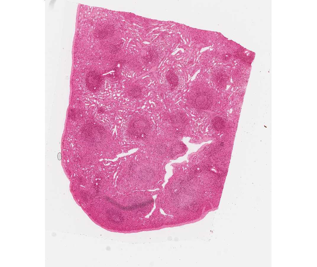 The spleen is comprised of red pulp and white pulp. The red pulp is the site of blood filtration and the white pulp is lymphoid tissue that responds to blood-borne antigens. Identify under low power some of the structures that are diagnostic of the organ. There is a dense connective tissue capsule that sends conspicuous trabeculae to partially subdivide the organ. Whereas the trabeculae derived from the capsule contain no vascular structures, those originated from the hilar region (which is not present in this section) carry a trabecular branch of the splenic artery and/or a trabecular tributary to the splenic vein. All the trabeculae contain some smooth muscle fibers. Unlike the thymus and lymph nodes, the spleen lacks division into cortex and medulla. Lymphatic nodules with or without germinal centers and with prominent eccentric central arterioles (called “central arteries) may be observed randomly distributed throughout the splenic pulp. These nodules together with areas of dense lymphatic tissue surrounding central arterioles, called the periarterial lymphatic sheaths (PALS), comprise the splenic white pulp. This lymphatic sheath is made up of T cells. Following stimulation, germinal centers containing B cells can be formed adjacent to the PALS, placing the "central arteriole" in an eccentric position. The remainder of the spleen consists of red pulp and is composed of sinusoids (modified blood vessels) and splenic cords (of Billroth). The latter are cellular regions organized as plates of loose lymphatic tissue separating the sinusoids.
It is not always possible to distinguish Billroth cords from the sinusoids easily, as is evident in this preparation where the sinusoids are partially collapsed after death. Under higher magnification, look for transverse and longitudinal sections of patent sinusoids. The lining cells of these sinusoids are elongated endothelial cells with tapered ends that lie parallel to the long axis of the vessel. These endothelial cells are separated from each other by gaps. In cross sections of sinusoids, therefore, the lining reticular cells are cut transversely and appear as cuboidal blocks arranged loosely in a circle, with intervening gaps. Look for the penicilli (short, straight arterioles that branch from the central artery and enter the red pulp). These penicilli branch into capillaries surrounded by accumulations of reticular cells and macrophages and known as "ellipsoids" (or "sheathed capillaries).
Between the white pulp and the red pulp is the marginal zone, a vascular region that is devoid of sinuses. It contains blood cells, lymphocytes, macrophages, and reticular cells. The region is the site of immunological activities due to the presence of numerous blood antigens.
#28 Spleen. H&E (not scanned)
This is a thicker section. Look for all the structures described above, noting particularly the central arteries, and penicillar arteries.
#25 Spleen, Monkey (Periodic acid-silver)
Open with WebViewer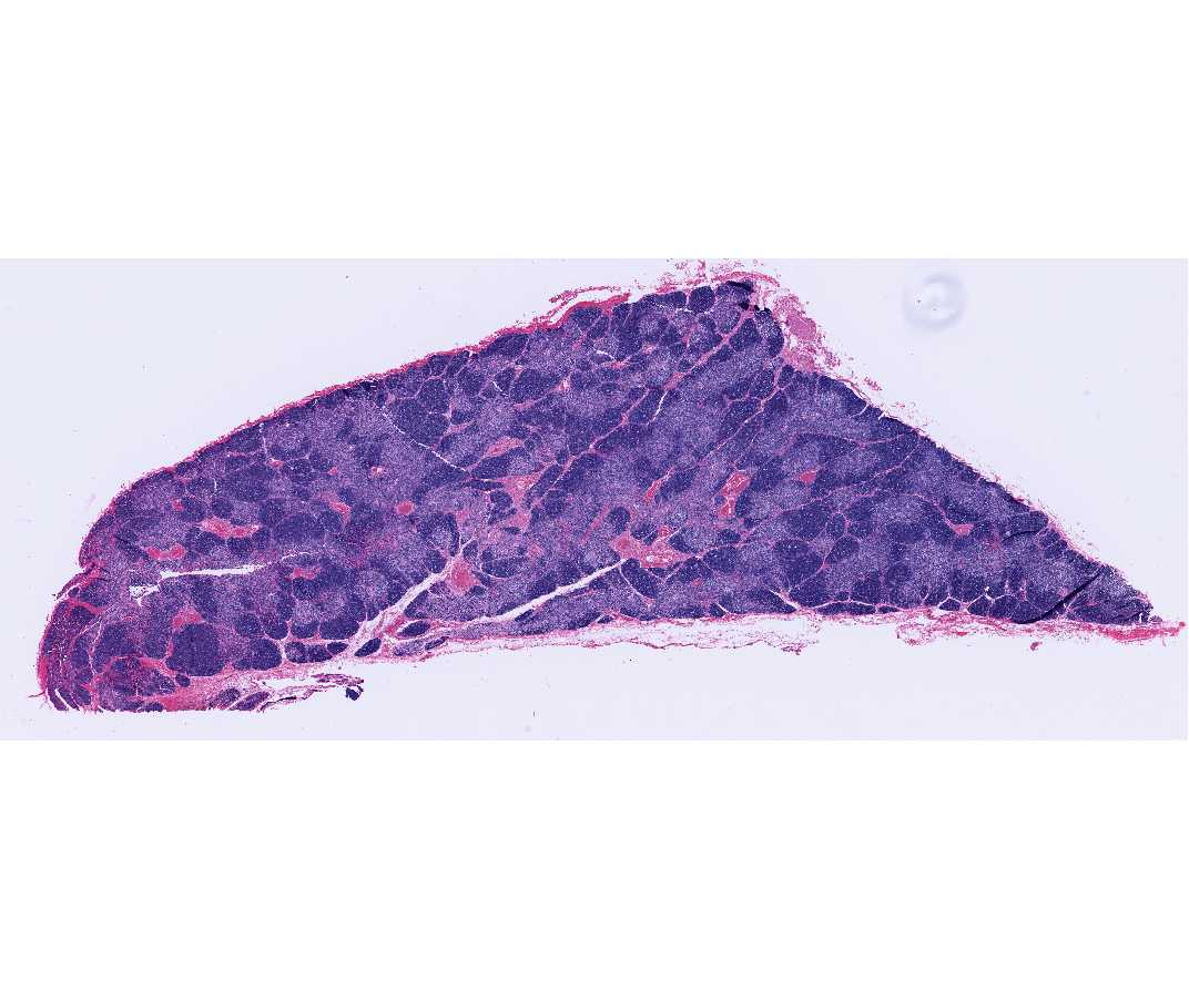
Like the PAS technique, this staining method, in addition to staining the network of reticular fibers, also stains the fenestrated basal laminae of the splenic sinusoids black. In section, the membrane may be seen as a succession of black points or short lines of silver-impregnated substance.
#30 Spleen, Monkey (Foot's silver and azo-carmine) (Not scanned)
This stain beautifully demonstrates the reticular fiber component of the capsule and trabeculae and the fine spongework of reticular fibers that form the delicate cytoskeleton of the splenic pulp (white pulp, marginal zone and red pulp). Note that the reticular fibers are thicker in the white pulp than in the remainder of the spleen and are continuous with those of the capsule and trabeculae. In the periarterial lymphatic sheaths of the white pulp, the meshwork of reticular fibers tends to follow a circumferential pathway about the central artery so that, in longitudinal sections of these sheaths, the fibers and their ensheathing reticular cells form parallel layers separated by lymphocytes. Notice that the reticular fibers are scarce or absent in the germinal centers of the white pulp nodules. The marginal zone and Billroth cords are permeated by a fine fibrous reticular meshwork. The sinusoids are surrounded by ring-like formations of reticular fibers.