SBPMD Histology Laboratory Manual
Joints
Bones are connected to one another at joints, or articulations. In gross anatomy, joints are usually classified according to the kind of movement they permit. An alternative classification is based upon the connective tissue structures that unite the bones entering into a joint. On such a morphological basis, four types of joints may be distinguished: (1) fibrous joints (syndesmoses), (2) cartilaginous joints (synchondroses), (3) osseous joints (synostoses), and (4) synovial joints (diarthrodial). Any of the first three types is an immovable or slightly movable joint. These are also called synarthroses because the articulating bones are kept together by the joint. Synovial joints are freely movable joints, because the articulating bones are kept apart by a synovial cavity. (These are also called diarthroses or diarthrodial joints.)
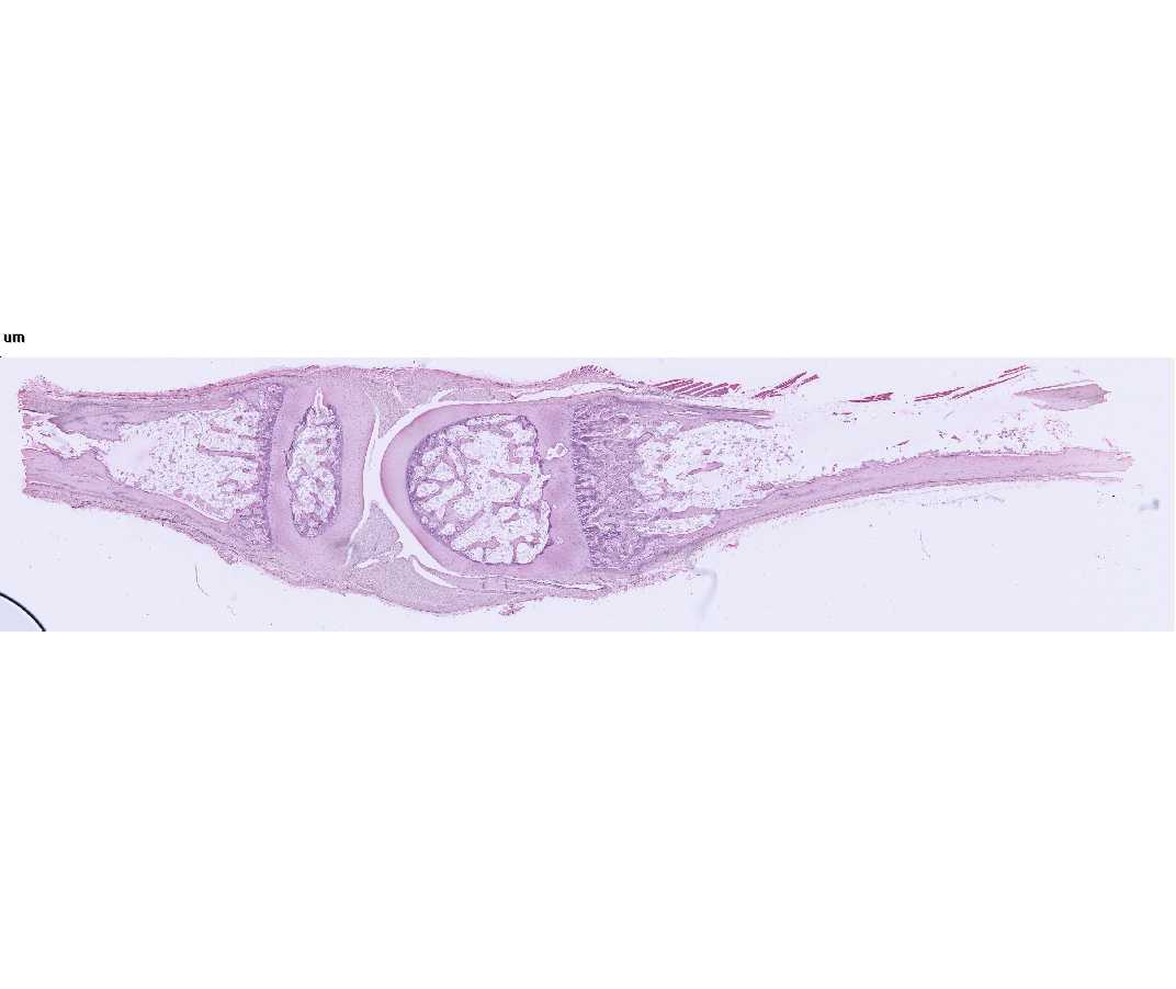
#95 or #97 Finger
Open with WebViewer
In synovial or diarthrodial joints, articular (hyaline) cartilage caps the ends of the bones, which are kept apart by a synovial cavity filled with synovial fluid, and the articulation is enclosed by a joint capsule. The latter is composed of an outer dense fibrous layer, the fibrous capsule. This is continuous with the periosteum over the bones and with an inner layer, the synovial membrane. The latter lines the joint cavity everywhere except over the articular cartilages. When present, fibrocartilaginous intra-articular disks or menisci are also lined by a synovial membrane. The synovial membrane is formed by a layer of collagenous fibers interspersed with flattened fibroblasts (synovial cells). This membrane may lie directly on the fibrous capsule or may be separated from it by connective tissue or fat. It is commonly thrown into folds (synovial villi) that project into the synovial cavity.
The synovial membrane is neither a membrane in the cell biological sense nor is it an epithelium. It is specialized, secretory connective tissue.
Locate with the naked eye the metacarpophalangeal joint and identify under low power the following structures: articular cartilages, synovial cavity, fibrous capsule, synovial membrane, and synovial villi. Then examine under high power the structure of the synovial membrane.
Questions
- What is the composition of synovial fluid? What are its functions?
- How are most of the cells of the articular cartilages nourished?