SBPMD Histology Laboratory Manual
Connective Tissue: Loose (Areolar)
This form of connective tissue has the largest number of cells per unit volume of extracellular matrix. The large number of cells frequently makes it difficult to distinguish the fibrous component without the use of special stains. The fibers in the matrix have a loose and irregular arrangement, and they consist of collagenous, elastic, or reticular fibers. Fibroblasts and macrophages are the most common cells in loose connective tissue, but mast cells, plasma cells, neutrophils and fat cells may also be found.
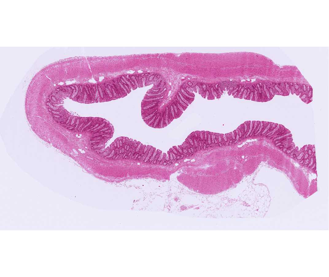
#39 Colon. Primate, H&E.
Open with WebViewer
Examine the slide with the scanning objective, and note that one surface is indented by pits that are lined by columnar epithelial cells. Immediately beneath these cells is the loose areolar connective tissue called lamina propria.
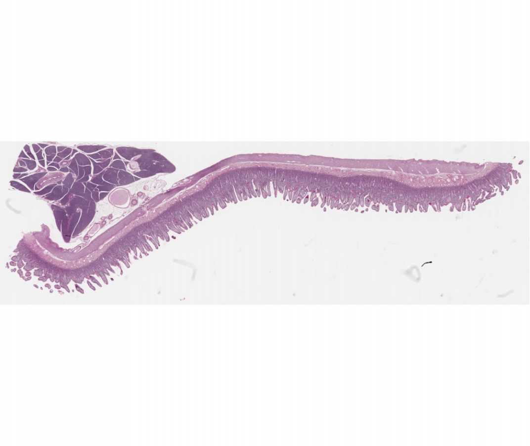
#101a Small Intestine. Guinea Pig, PAS and hematoxylin
Open with WebViewer
This section stained with PAS allows one to identify the numerous macrophages laden with large pink granules (lysosomes) located in the lamina propria.