SBPMD Histology Laboratory Manual
Cells, Organelles: Basic and Acid Stains
The first step in preparing a tissue or organ for microscopic examination is fixation, or preservation, of the specimen. Formalin is a commonly used fixative. Many other fixatives are available and are used in the study of specific structures.
Note: There is a more complete description of methods for preparation of histological samples at the end of this laboratory manual (p. 105) under the heading “Technique”.
The specimen on the microscope slide is a thin slice, or section, of the fixed tissue or organ. The section is stained by one or more dyes. Without staining the section would be nearly invisible with the microscope. Components of the specimen generally stain selectively and, on this basis, various regions of the specimen may be differentiated from each other.
Most dyes are neutral salts. In some stains, the dye moiety is a cation and such dyes are called cationic or basic dyes. These form salts with tissue anions, especially the phosphate groups of the nucleic acids and the sulfate groups of the glycosaminoglycans. When the dye moiety is an anion, the dye is called anionic or acid dye and salt formation occurs with tissue cations including the lysine and arginine groups of tissue proteins. Tissue components that recognize basic dyes are "basophilic" and those that recognize acid dyes are "acidophilic". A common combination of stains is hematoxylin and eosin (H&E), which are commonly referred to as basic and acid dyes, respectively.
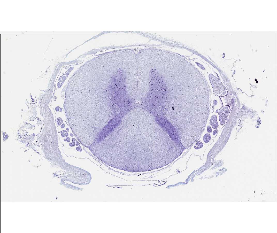
#85 Spinal cord. Nissl stain
Open with WebViewer
Look at the slide with the naked eye ("transilluminate" the slide) and with the scanning objective of your microscope. Note the butterfly-shaped, blue region in the center of the section, which is called the gray matter of the spinal cord. Scattered in the gray matter are cell bodies of neurons (nerve cells). These cells are among the largest in the body and because of their size and staining characteristics serve as a useful introduction to the study of cellular structure. Examine several neurons with the 10x objective. Identify the nucleus, the prominent nucleolus within the pale staining nucleus, and the cytoplasm. Locate a neuron with a distinct nucleus and nucleolus and study it with the 40x objective. Examine selected areas using the oil immersion objective. Ask an instructor for help if you are unfamiliar with the use of this objective. (Remember not to get oil on the 40x objective.) Note the basophilic bodies scattered in the cytoplasm. These darkly stained particles are called Nissl substance (or bodies) and represent stacks of the rough endoplasmic reticulum and free polyribosomes. What is the rough endoplasmic reticulum and with what synthetic activity is it associated? Why are the nucleus and the nucleolus not visible in some neurons?
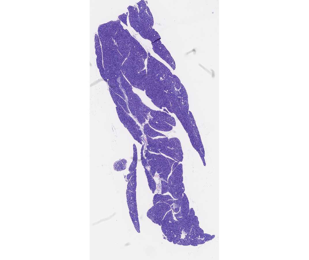
#107 Pancreas. Guinea pig. Acid fuchsin-toluidine blue
Open with WebViewer
After observing this exocrine gland at low power, examine the cells at 40x. Identify the base and apex of the cells of a secretory unit, the acinus. Note the basophilia in the basal compartment and the acidophilia in the apical (luminal) compartment of the cytoplasm. What subcellular organelle is responsible for attracting the basic stain? How do you account for the acidophilia in the apical compartment?
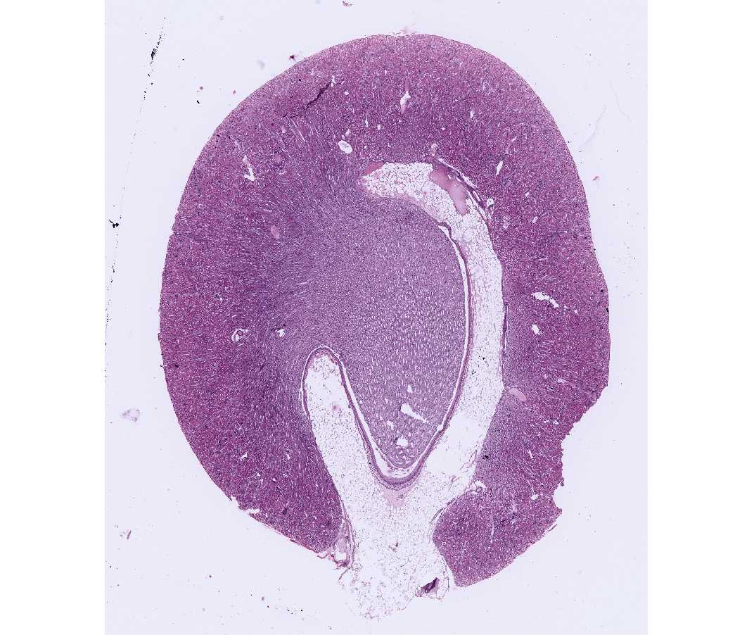
#50 Kidney. Guinea pig. H&E
Open with WebViewer
This slide illustrates different kinds of cells; do not be concerned at this time with the structure of the kidney. Large numbers of cells are seen. Nuclei are basophilic and are stained blue. At lower magnifications they appear as blue dots and at higher magnifications chromatin and nucleoli may be identified within the nucleus. Surrounding the nucleus is the acidophilic cytoplasm stained pink (due to the positive charges on arginine and lysine). Many cells of the kidney tubules appear confluent because the cell (plasma) membranes are not stained by this technique; do not confuse the lumen of the tubule with a cell. The free surface of the cell, facing a lumen, is referred to as the cell apex and the opposite surface is the cell base.